Keratoconus is an eye disease that occurs when the cornea loses its shape, thins and becomes conical. This prevents light from focusing correctly and causes vision problems.

Prof. Dr. Aylin Kılıç Eye Diseases and Surgery
Keratoconus is an eye disease that occurs when the cornea loses its shape, thins and becomes conical. This prevents light from focusing correctly and causes vision problems.
Prof. Dr. Aylin Kılıç played a critical role in the design and development of KeraNatural, an innovative approach to the treatment of keratoconus. As the first female ophthalmologist invited to collaborate by “VisionGift,” a leading eye bank in the United States, she earned the title “Mother of KeraNatural.”


Keratoconus is a progressive eye condition that arises when the shape of the cornea becomes distorted, directly causing impaired vision.
Depending on the degree of the disease, there may be cases in which the patient’s vision is almost lost. Intervention and a treatment plan are needed before serious vision loss occurs.
The cornea is the outermost layer of the eye, located at the very front: a transparent layer with a regular dome shape. Important for vision, this layer ensures that light is focused correctly in a healthy eye. The answer to the question what is keratoconus also lies in the cornea.
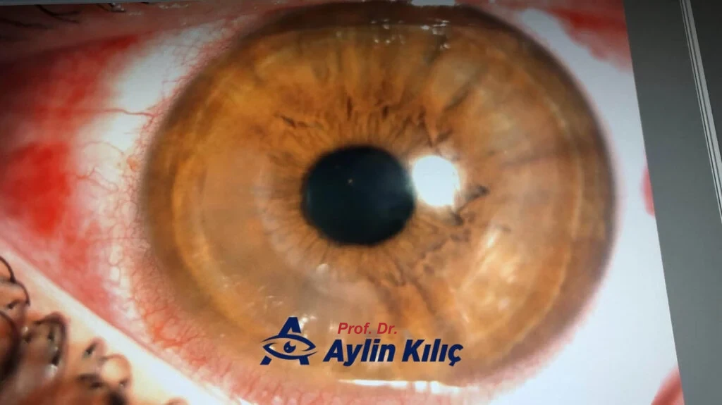
Keratoconus is an eye disease that occurs when the regular dome shape of the cornea is disrupted and it becomes pointed and bulges forward. Because of this deformity in the cornea, light entering the eye cannot be focused correctly on the visual center. The patient’s vision begins to deteriorate according to the degree of this deformity.
The result is blurred vision and distorted vision that increases over time. The patient experiences noticeable vision problems in activities such as driving or reading.
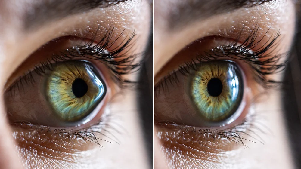
Although there is a clear answer to the question what is keratoconus, it is not possible to give a definitive answer to the question what causes keratoconus.
Scientific research has not been sufficient to reach a firm conclusion about the causes of the disease. Nevertheless, certain factors stand out in the research as possible causes of the disease. Let us give more information about what may cause the disease through these factors.

The biggest cause of keratoconus is rubbing the eyes. Rubbing the eyes because of itching or for any other reason places a certain amount of pressure on the cornea. This pressure is the biggest cause of the cornea losing its shape.
For this reason, patients with dry eye in particular need to avoid scratching or rubbing their eyes and to get treatment for the dry eye that creates this sensation.
All of these can be cited among the causes of keratoconus, but let us remind you once again that the exact causes have not yet been determined or scientifically proven.
In keratoconus treatment, there are different treatment options chosen according to the progression and current state of the disease. Here we should talk about the treatments that deliver the most effective results.
One of the new-generation treatments, corneal cross-linking (CXL) is a treatment that aims to strengthen the cornea and thereby minimize the effects of keratoconus.
Corneal cross-linking involves the use of ultraviolet (UV) light and riboflavin (vitamin B2) eye drops. When exposed to UV light, riboflavin forms chemical bonds within the cornea, increasing its structural integrity and further strengthening it. This process effectively halts the progression of keratoconus and can even improve the shape of the cornea.
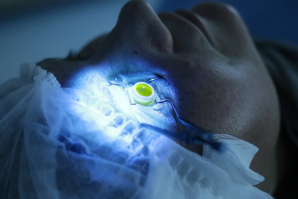
The CXL procedure is typically an outpatient procedure lasting about an hour. After the eye is numbed with local anesthetic eye drops, the eye doctor applies riboflavin drops to the cornea. The eye is then exposed to UV light. Patients may experience temporary discomfort during the procedure, but the benefits far outweigh the momentary discomfort.
After CXL, patients may feel sensitivity to light and mild discomfort for a few days. Protective lenses are generally recommended to protect the eyes during this initial recovery period. Most people can resume their normal activities within a week, and gradual improvements in vision become apparent over the following months.
Developed after extensive clinical research, KeraNatural – CAIRS treatment focuses on the underlying causes of the disease and aims directly to improve the patient’s vision.
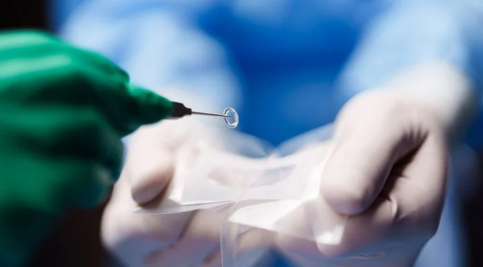
KeraNatural, or the allograft corneal ring, is the name given to a semicircular piece of collagen/cornea used to improve the patient’s vision.
The safe corneal tissue, which is human collagen, is packaged in an entirely safe manner, sterilized and made ready for use at the Visiongift Eye Bank in the United States.
In the procedure, this ring obtained from human collagen is implanted into the patient’s cornea, where it is effective in flattening and thickening the cornea.
KeraNatural, placed inside a tunnel created in the cornea using a laser, is among the most effective methods for improving the patient’s vision.
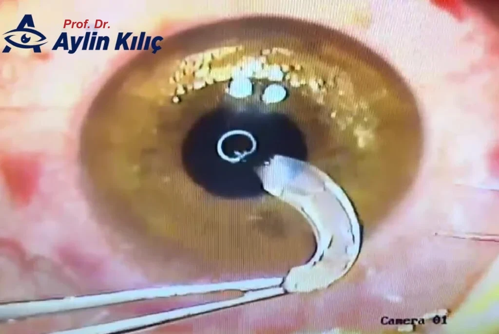
In short, KeraNatural is a minimally invasive and controlled procedure that aims to improve vision for individuals with corneal conditions, keratoconus in particular. It offers the advantage of faster recovery, less discomfort and the ability to adjust the treatment as needed for optimal results.
However, individual responses to the procedure may vary. It is very important that patients discuss their particular circumstances, expectations and any concerns with their eye care professionals before undergoing CAIRS.
| PMMA rings | Alternative fresh tissue for CAIRS | KeraNatural® | |
|---|---|---|---|
| Reversible and adjustable procedure | Yes | Yes | Yes |
| Efficacy for improving vision | High | High | High |
| Sterilized, tested, and preserved | Yes | No (donor tissue quality and infection risk vary) | Yes |
| Risk of extrusion | Significant | Minimal | Minimal |
| Biocompatibility | Low | High | High |
| Product supply and availability | Continuous supply and availability | Limited supply, waiting lists | Continuous supply and availability |
| Logistics (storage and shelf life) | 5-year shelf life at room temperature | 2 weeks to get tissue from donor to patient under refrigerated sterile conditions | Simple: 2-year shelf life at room temperature |
| Pre-cut segments | Yes | No, the surgeon must cut the arc from the donor cornea | Yes |
| Suitable for patients with thin corneas | No, deep incision | Yes | Yes |
Which treatment is chosen and what kind of treatment process is followed must always be decided together with a specialist doctor. We should also say that the doctor’s experience and skills are very important here. A roadmap for treatment will be drawn up after a detailed examination.
In addition, after the chosen operation the patient must follow all of the doctor's recommendations and avoid rubbing their eyes. As we said at the beginning, eye rubbing is one of the most important factors in keratoconus and can result in the patient experiencing vision problems again after treatment.
It should also be remembered that this is a progressive disease. Regular eye examinations are also important for early diagnosis and treatment.
You can contact me about your condition, my treatment methods and anything else you would like to know.
Keratoconus eye disease is a progressive vision disorder that occurs when the cornea becomes thinner and more pointed than normal. Usually beginning at a young age, this disease lowers the quality of vision over time and can cause complaints of blurred vision that glasses do not fully correct.
Today, thanks to early diagnosis and advanced technologies, keratoconus treatment delivers quite successful results. In particular, CCL X treatment (corneal cross-linking, CXL) is one of the most effective methods for halting the progression of the disease by strengthening the corneal tissue.
In advanced-stage cases, keratoconus surgery may be preferred. This surgical procedure is planned according to the patient's corneal structure, and special lenses or rings may be used to improve visual acuity.
Among the best keratoconus doctors, Prof. Dr. Aylin Kılıç offers each patient a personalized treatment plan with modern diagnostic devices and her experienced team. When keratoconus eye disease is diagnosed early and treated with the right methods, the risk of vision loss can be minimized.
If you too are experiencing symptoms such as blurred or double vision, you can have an eye examination without delay and seek support from a specialist physician for keratoconus treatment.
Keratoconus is an eye disease that occurs when the cornea loses its shape, thins and becomes conical. This prevents light from focusing correctly and causes vision problems.
Yes, keratoconus can be treated with different methods. Treatment options include corneal cross-linking (CXL), KeraNatural (CAIRS) and corneal transplant. The treatment method is determined according to the stage of the disease and the condition of the cornea.
Keratoconus treatment prices vary depending on the treatment method chosen, the technology used and the clinic providing the treatment. For example:
Corneal Transplant: Because it is a more advanced procedure, prices may be $5,000 and above.
For detailed pricing information, we recommend contacting your doctor or the relevant clinics.
With many experienced doctors and modern clinics specializing in eye health, Istanbul is one of Türkiye’s leading cities in this field. Prof. Dr. Aylin Kılıç is one of the specialists with international experience in this field.
Keratoconus treatment should be performed by ophthalmologists who specialize in corneal surgery. Points to consider when choosing a doctor:
Yes, with early diagnosis and the right treatment, the progression of keratoconus can be stopped and serious vision loss can be prevented. However, if left untreated, the disease can worsen vision problems.
Because local anesthesia is used during treatment, pain is generally not felt. There may be mild discomfort after treatment, but this is short-lived.
The duration varies according to the treatment method and the patient's condition:
Corneal Transplant: It requires a longer time and recovery period.
Although the exact causes of keratoconus are not fully known, genetic predisposition is an important factor. Individuals with a family history are thought to be potentially at risk.
 Does Vision Increase with CAIRS Technique in Keratoconus Treatment?
Read more
 Can Phakic Intraocular Lens Be Applied to Patients with Keratoconus?
Read more
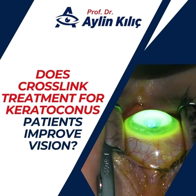 Does Crosslink Treatment for Keratoconus Patients Improve Vision?
Read more
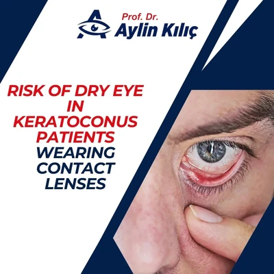 Risk of Dry Eye in Keratoconus Patients Wearing Contact Lenses
Read more
 Is Wearing Glasses Compulsory in Keratoconus?
Read more
 Can Keratoconus Occur in Children? Is Early Diagnosis Important?
Read more
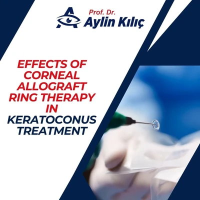 Effects of Corneal Allograft Ring Therapy in Keratoconus Treatment
Read more
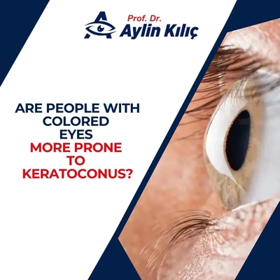 Are People with Colored Eyes More Prone to Keratoconus?
Read more